
C57BL/6-Cd3etm1(CD3E)Bcgen Cd3dtm1(CD3D)Bcgen Cd3gtm1(CD3G)Bcgen B2mtm1(B2M/HLA-A2.1/H2-D)Bcgen Fcgrttm1(B2m/Fcgrt)Bcgen/Bcgen • 114542
| Product name | B-hCD3EDG/HLA-A2.1 plus mice |
|---|---|
| Catalog number | 114542 |
| Strain name | C57BL/6-Cd3etm1(CD3E)Bcgen Cd3dtm1(CD3D)Bcgen Cd3gtm1(CD3G)Bcgen B2mtm1(B2M/HLA-A2.1/H2-D)Bcgen Fcgrttm1(B2m/Fcgrt)Bcgen/Bcgen |
| Strain background | C57BL/6 |
| NCBI gene ID | (Human) |
| Aliases | T3E; TCRE; IMD18; CD3epsilon; T3D; IMD19; CD3DELTA; CD3-DELTA; T3G; IMD17; CD3GAMMA; CD3-GAMMA; HLAA; IMD43; AMYLD6; MHC1D4; FCRN; FcgammaRn; alpha-chain |
Gene targeting strategy for B-hCD3EDG/HLA-A2.1 plus mice.
The chimeric human CD3EDG was expressed, while mouse Cd3edg were knocked out in B-hCD3EDG/HLA-A2.1 plus mice.
The B2M gene (Exon1 to Exon3) of mouse were replaced by the sequence encompassing the human B2M CDS and HLA-A*0201 gene that included leader sequence, α1 and α2 domains ligated to a fragment of the murine H-2Db gene containing the α3, transmembrane and cytoplasmic domains in B-hCD3EDG/HLA-A2.1 plus mice.
The mouse B2M gene is knocked into exon 2 of the mouse Fcgrt gene and is fused via a linker to the remaining portion of exon 2, a strategy that enables the co-expression of mouse B2M and Fcgrt in B-hCD3EDG/HLA-A2.1 plus mice.
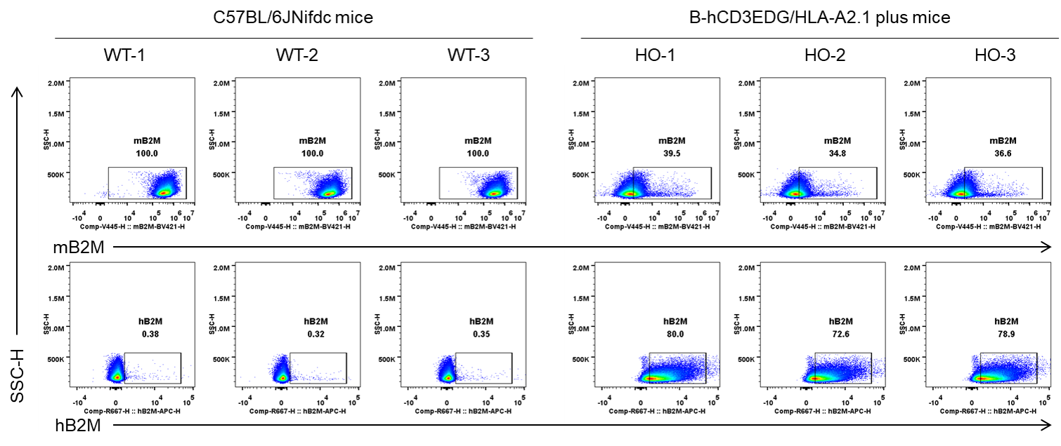
Strain specific B2M expression analysis in wild-type mice (WT) and homozygous (HO) B-hCD3EDG/HLA-A2.1 plus mice by flow cytometry. Splenocytes were collected from wild-type (WT) C57BL/6JNifdc mice and homozygous (HO) B-hCD3EDG/HLA-A2.1 plus mice (female, 8-week-old, n=3). Protein expression was analyzed with anti-mouse B2M antibody (BD Biosesciences, 744802), and anti-hB2M antibody (Biolegend, 395712) by flow cytometry. Mouse B2M was detectable in both C57BL/6JNifdc mice and B-hCD3EDG/HLA-A2.1 plus mice because the mouse B2M gene is knocked into exon 2 of the mouse Fcgrt gene and is fused via a linker to the remaining portion of exon 2, a strategy that enables the co-expression of mouse B2M and Fcgrt in B-hCD3EDG/HLA-A2.1 plus mice. Human B2M was only detectable in B-hCD3EDG/HLA-A2.1 plus mice.
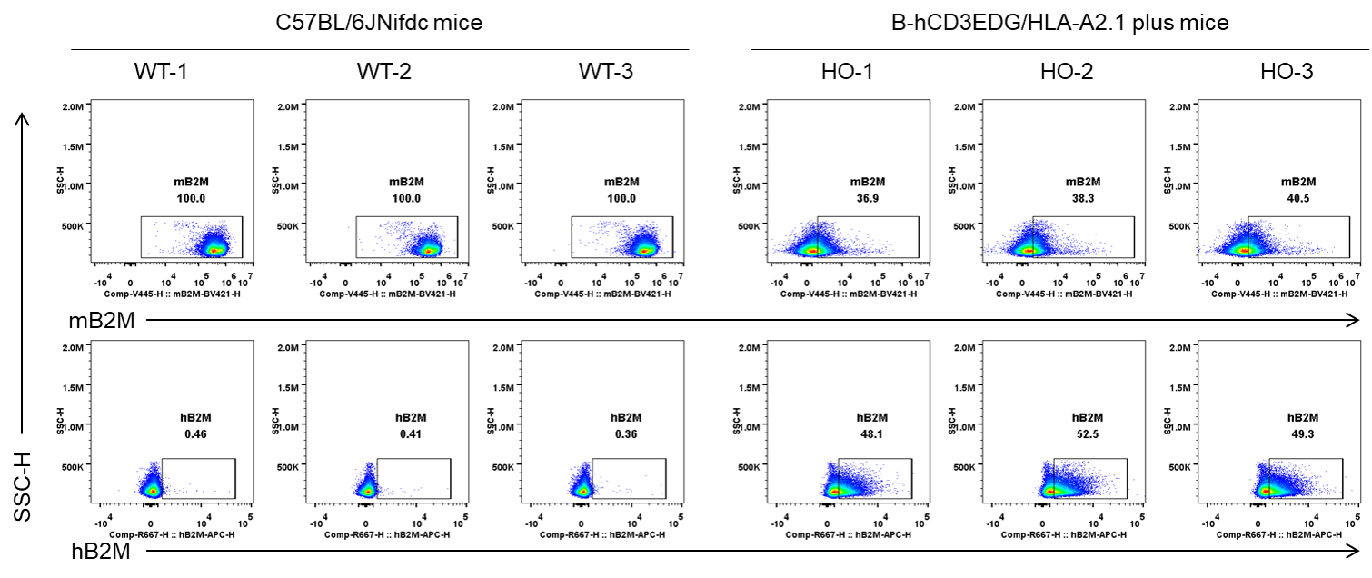
Strain specific B2M expression analysis in wild-type mice (WT) and homozygous (HO) B-hCD3EDG/HLA-A2.1 plus mice by flow cytometry. Blood cells were collected from wild-type (WT) C57BL/6JNifdc mice and homozygous (HO) B-hCD3EDG/HLA-A2.1 plus mice (female, 8-week-old, n=3). Protein expression was analyzed with anti-mouse B2M antibody (BD Biosesciences, 744802), and anti-hB2M antibody (Biolegend, 395712) by flow cytometry. Mouse B2M was detectable in both C57BL/6JNifdc mice and B-hCD3EDG/HLA-A2.1 plus mice because the mouse B2M gene is knocked into exon 2 of the mouse Fcgrt gene and is fused via a linker to the remaining portion of exon 2, a strategy that enables the co-expression of mouse B2M and Fcgrt in B-hCD3EDG/HLA-A2.1 plus mice. Human B2M was only detectable in B-hCD3EDG/HLA-A2.1 plus mice.
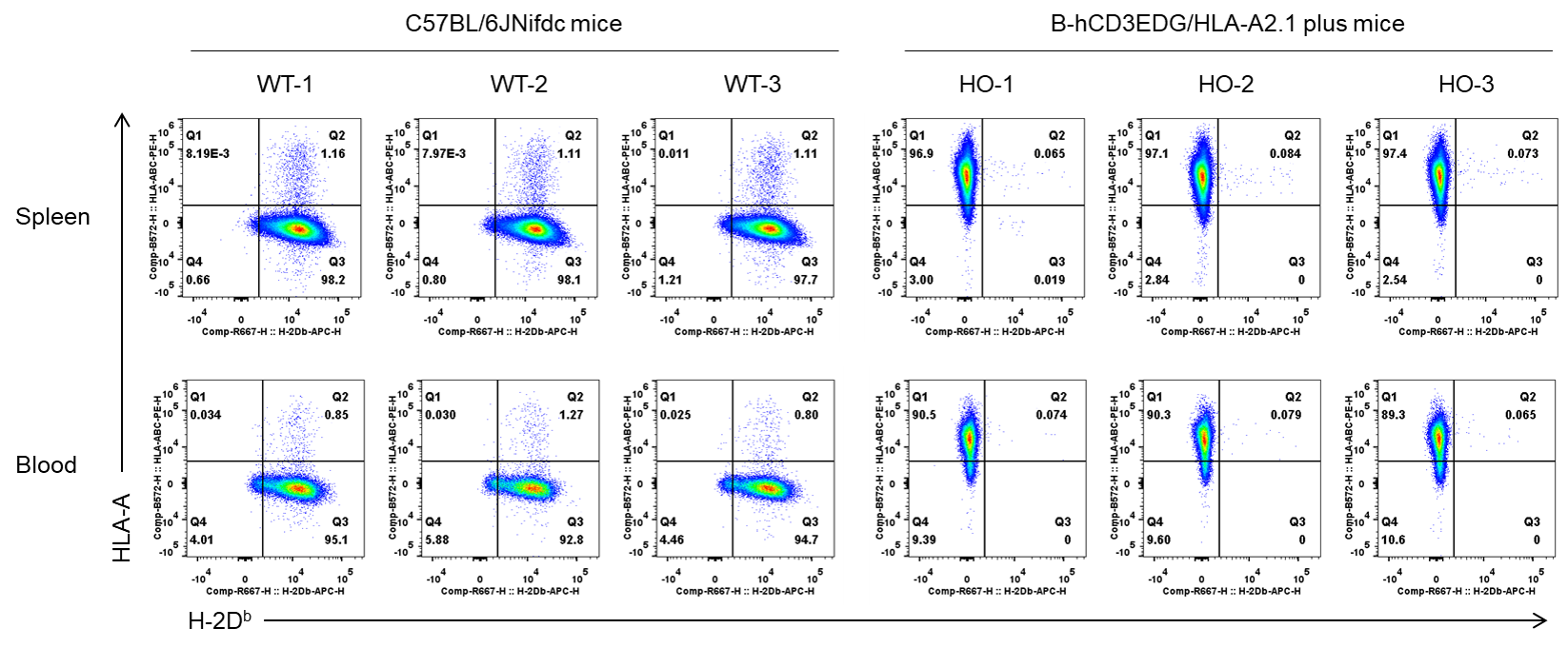
Strain specific B2M expression analysis in wild-type mice (WT) and homozygous (HO) B-hCD3EDG/HLA-A2.1 plus mice by flow cytometry. Splenocytes and blood cells were collected from wild-type (WT) C57BL/6JNifdc mice and homozygous (HO) B-hCD3EDG/HLA-A2.1 plus mice (female, 8-week-old, n=3). Protein expression was analyzed with anti-H-2Db antibody (Biolegend, 111513) and anti-HLA-ABC antibody (Biolegend, 311406) by flow cytometry. Mouse H-2Db was detectable in C57BL/6JNifdc mice. Human HLA-A was only detectable in B-hCD3EDG/HLA-A2.1 plus mice but not in wild-type C57BL/6JNifdc.
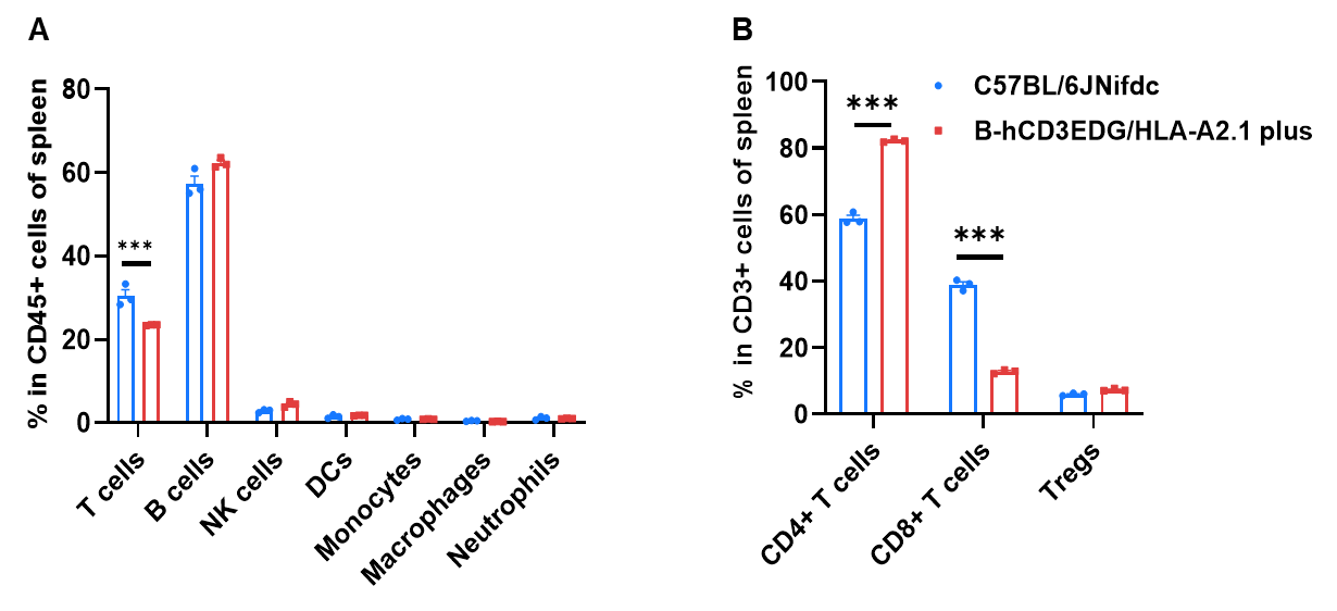
Frequency of leukocyte subpopulations in spleen by flow cytometry. Splenocytes were isolated from wild-type C57BL/6JNifdc mice and homozygous B-hCD3EDG/HLA-A2.1 plus mice (female, 8-week-old, n=3). A. Flow cytometry analysis of the splenocytes was performed to assess the frequency of leukocyte subpopulations. B. Frequency of T cell subpopulations. Frequencies of B cells, NK cells, dendritic cells, monocytes, macrophages, neutrophils, and Tregs in B-hCD3EDG/HLA-A2.1 plus mice were similar to those in C57BL/6JNifdc mice. Frequencies of T cells and CD8+ T cells in B-hCD3EDG/HLA-A2.1 plus mice were lower than that in C57BL/6JNifdc mice, whereas the frequency of CD4+ T cells in B-hCD3EDG/HLA-A2.1 plus mice was higher than that in C57BL/6JNifdc mice. Values are expressed as mean ± SEM. Significance was determined by two-way ANOVA test. *P < 0.05, **P < 0.01, ***p < 0.001.
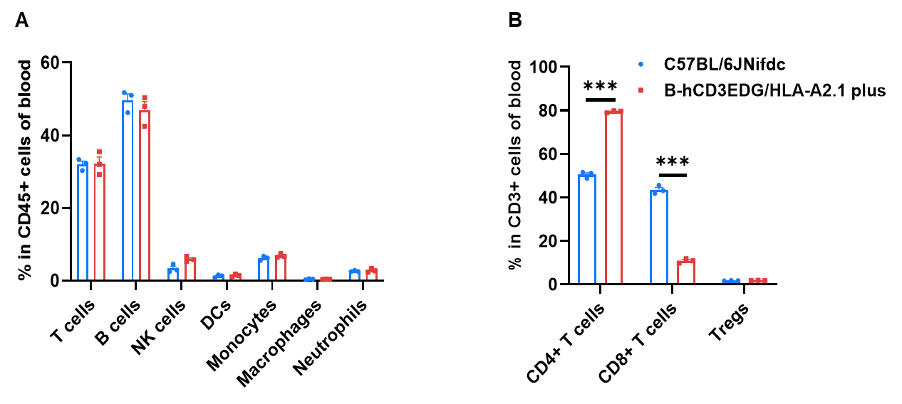
Frequency of leukocyte subpopulations in blood by flow cytometry. Blood cells were isolated from wild-type C57BL/6JNifdc mice and homozygous B-hCD3EDG/HLA-A2.1 plus mice (female, 8-week-old, n=3). A. Flow cytometry analysis of the blood was performed to assess the frequency of leukocyte subpopulations. B. Frequency of T cell subpopulations. Frequencies of T cells, B cells, NK cells, dendritic cells, monocytes, macrophages, neutrophils, and Tregs in B-hCD3EDG/HLA-A2.1 plus mice were similar to those in C57BL/6JNifdc mice. Frequency of CD8+ T cells in B-hCD3EDG/HLA-A2.1 plus mice were lower than that in C57BL/6JNifdc mice, whereas the frequency of CD4+ T cells in B-hCD3EDG/HLA-A2.1 plus mice was higher than that in C57BL/6JNifdc mice. Values are expressed as mean ± SEM. Significance was determined by two-way ANOVA test. *P < 0.05, **P < 0.01, ***p < 0.001.
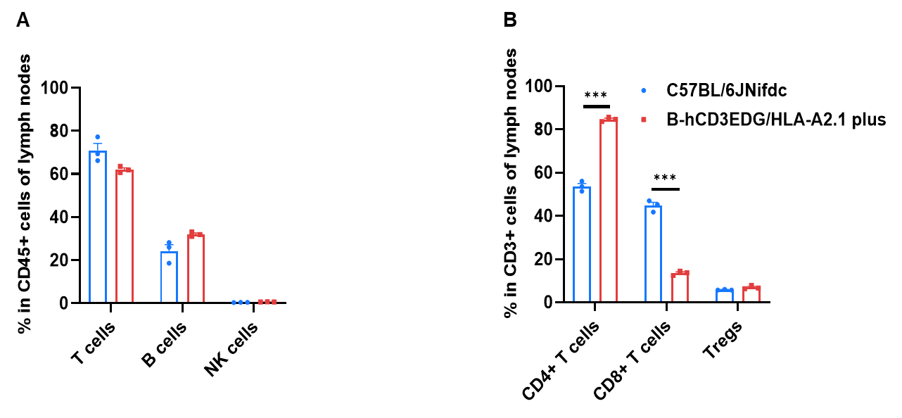
Frequency of leukocyte subpopulations in lymph node by flow cytometry. Lymph node cells were isolated from wild-type C57BL/6JNifdc mice and homozygous B-hCD3EDG/HLA-A2.1 plus mice (female, 8-week-old, n=3). A. Flow cytometry analysis of the lymph node was performed to assess the frequency of leukocyte subpopulations. B. Frequency of T cell subpopulations. Frequencies of T cells, B cells, and NK cells in B-hCD3EDG/HLA-A2.1 plus mice were similar to those in C57BL/6JNifdc mice. Frequency of CD8+ T cells in B-hCD3EDG/HLA-A2.1 plus mice were lower than that in C57BL/6JNifdc mice, whereas the frequency of CD4+ T cells in B-hCD3EDG/HLA-A2.1 plus mice was higher than that in C57BL/6JNifdc mice. Values are expressed as mean ± SEM. Significance was determined by two-way ANOVA test. *P < 0.05, **P < 0.01, ***p < 0.001.
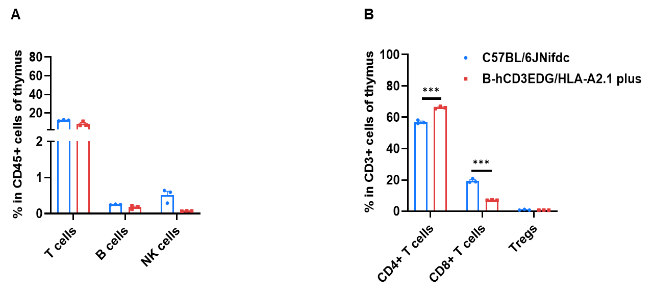
Frequency of leukocyte subpopulations in lymph node by flow cytometry. Thymocytes were isolated from wild-type C57BL/6JNifdc mice and homozygous B-hCD3EDG/HLA-A2.1 plus mice (female, 8-week-old, n=3). A. Flow cytometry analysis of the lymph node was performed to assess the frequency of leukocyte subpopulations. B. Frequency of T cell subpopulations. Frequencies of T cells, B cells, and NK cells in B-hCD3EDG/HLA-A2.1 plus mice were similar to those in C57BL/6JNifdc mice. Frequency of CD8+ T cells in B-hCD3EDG/HLA-A2.1 plus mice were lower than that in C57BL/6JNifdc mice, whereas the frequency of CD4+ T cells in B-hCD3EDG/HLA-A2.1 plus mice was higher than that in C57BL/6JNifdc mice. Values are expressed as mean ± SEM. Significance was determined by two-way ANOVA test. *P < 0.05, **P < 0.01, ***p < 0.001.